3D/4D Ultralyd
3D-ultralyd gjør at en ser bredde, høyde og dybde i bildet, men en ser ikke bevegelse. 4-dimensjonal (4D) ultralyd viser fosterets bevegelser i 3D-bilder( film snut).
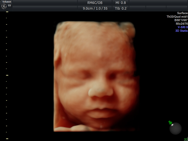
3D-avbilding av foster er blitt en populær og etterspurt teknologi fra den gravide . For å få gode bilder i 3D/4D er det best å ta denne undersøkelsen mellom 24-32 uker. Den beste tiden for 3D er uke 28. Det er mulig å prøve å ta disse bildene i andre uker også.
Om du vet at du har morkaken i fremre vegg, har du større sjanser at få bra bilder ved 25-26 uker.
Det er alltid en hyggelig begivenhet med tvillinger. Si fra om dette når du bestiller timen. Tvillinger avbildes best mellom 23-28 uker.
Når barnet ligger litt vanskelig til for å få tatt gode bilder prøver vi med litt saft eller annet søtt til mor. Det er lurt å ha spist før en kommer. Vi setter av god tid til undersøkelsen slik at alle betingelser er til stede for at få tatt gode bilder.
En får vanligvis sett barnet sitt på denne måten etter svangerskapsuke 24, og før uke 36, men dette kan variere.