 3D / 4D Ultrasound
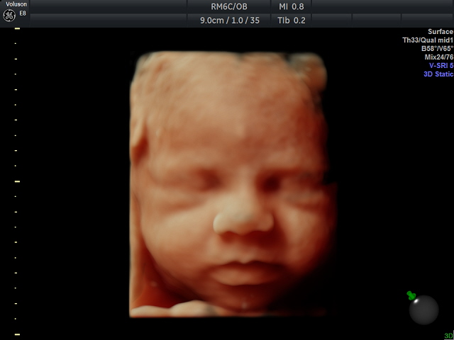
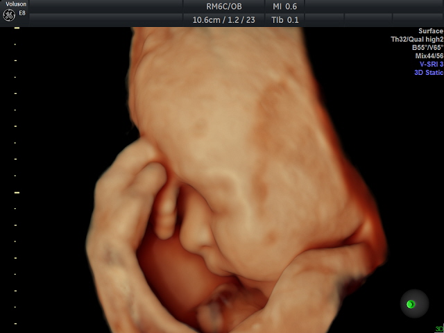
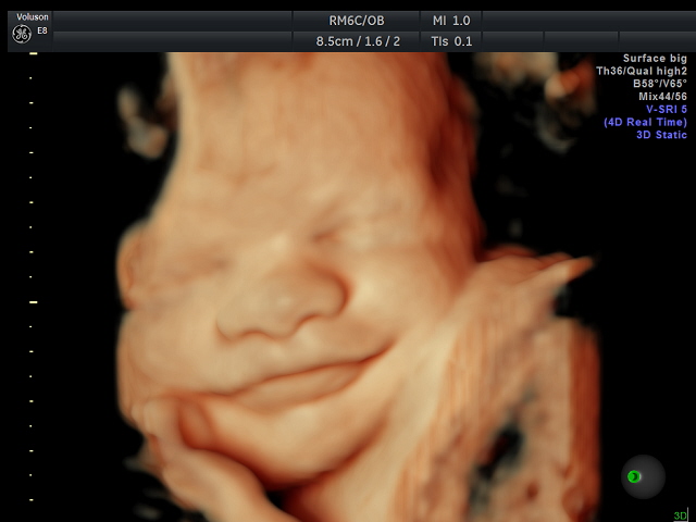
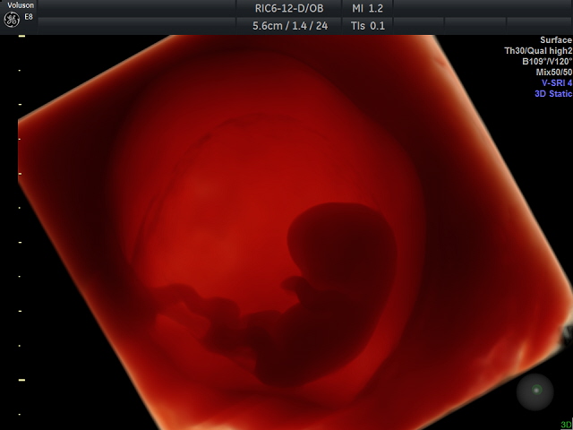
3D ultrasound allows one to see the width, height and depth of the picture, but there is no movement. 4-dimensional (4D) ultrasound shows fetal movement in 3D images.
3D imaging of fetuses has become a popular and sought-after technology by pregnant women. It is possible to take good 3D/4D pictures any time throughout the pregnancy, but the optimal time is to take them between 25-30 weeks.
If you know you have placenta in the anterior wall, you have a greater chance to get good images at 25 to 26 weeks.
Being pregnant with twins is always especially exciting. Let us know if you are pregnant with twins when you call to make an appointment as the pictures of twins are best to be taken between 23-28 weeks.
When the position of your baby is not optimal for taking the pictures, we try giving some juice or something sweet to the Mother. It is advisable to have eaten something before arriving. We set aside plenty of time for the examination so that all the conditions are optimal for good pictures. For 3D/4D ultrasounds, we use about 60 minutes for the entire examination. If you have had any previous examinations then it may take less time.
One can usually see their baby this way after 24 weeks of gestation and until week 36, though it can vary.
3D Ultralyd
3D Ultralyd
3D Ultralyd
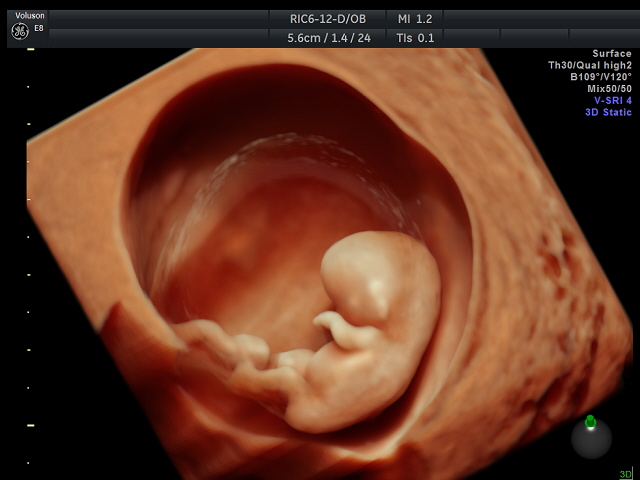
Tidlig 3D Ultralyd
Tidlig 3D Ultralyd

Tidlig 3D Ultralyd
Copyright GE Vingmed Ultrasound AS / Images made by Dr. Benoit